NL-GHK-Cu peptide therapy, owing to the complex mechanism of action discussed in the article below, enables increased expression of epidermal stem-cell markers such as integrins and p63, which in turn supports the elimination of problems associated with hyperkeratosis and excessive skin thickening.
Abstract: Once the NL-GHK-Cu peptide enters the body, biological processes are initiated that promote the expression of epidermal stem-cell markers, thereby inhibiting the development of—and contributing to the elimination of—hyperkeratotic skin lesions of diverse etiologies.
Keywords: NL–GHK-Cu; skin structure; skin functions; epidermis; epidermal layers; thickening; keratinization; stem cells; integrins
Introduction
Studies investigating the efficacy of NL-GHK-Cu have indicated that this peptide may contribute to the reduction of hyperkeratosis and skin thickening arising from, among others, genetic and environmental factors, and may also support prophylactic therapy aimed at preventing their development.
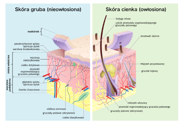
Skin Structure
As is well established, the skin has a highly complex structure and, as the body’s outer covering, it is considered one of the largest organs. The condition of the skin often reflects the overall condition of the organism. The skin consists of two primary layers: an external layer and an internal layer. The first, visible, hair-bearing external layer is the epidermis, which forms a barrier between the external environment and the body’s internal milieu. The second, non-visible, non–hair-bearing layer is the dermis, which contains important nerve endings, hair follicles, and sweat and sebaceous glands.
The subcutaneous layer, although closely associated with the skin, is not considered a structural component of it. It is composed predominantly of adipose tissue, with only a small proportion consisting of connective tissue. The subcutaneous tissue contains nerve fibers and blood vessels. Its principal role is participation in thermoregulation, which is of major importance for the organism as a whole.
Skin Functions
Human skin performs numerous physiological functions. By fulfilling an essential protective role, it shields internal organs from harmful environmental, physical, chemical, and microbiological factors. Through these functions, the skin helps maintain homeostasis between the organism and its surroundings. The most widely recognized functions include barrier protection, thermoregulation, contributions to water balance and secretory processes, participation in protein synthesis and the synthesis of selected compounds, and involvement in the metabolism of proteins, lipids, and carbohydrates. In addition, the skin plays an important role in immune responses and in the conduction of sensory stimuli. Healthy skin is free from damage related to improper care or adverse environmental impact, tolerates changes in ambient temperature and humidity, and responds appropriately to most tested and properly formulated skincare products.
Epidermis
The epidermis is composed almost entirely of living cells—keratinocytes—which undergo keratinization, i.e., transformation into dead, dry, anucleate corneocytes as they migrate upward.
Layers of the epidermis:
Basal (germinative) layer: a layer of stem cells with an almost unlimited capacity for cell division, supplying new cells to the surface.
Spinous layer: cells in this layer are not capable of division or growth; the spinous layer is thicker in men and becomes thinner with age in both sexes.
The basal and spinous layers constitute the so-called living epidermis (the Malpighian layer).
Granular layer: the second stage of cellular differentiation; the cell nucleus disintegrates and the process of forming rigid keratin fibers begins.
Stratum corneum: the final stage of differentiation; the young keratinocyte becomes a hard, water-resistant cell—i.e., a corneocyte.
Skin Keratinization
The cause of hyperkeratosis—i.e., excessive keratinization—is overly rapid proliferation of epidermal cells, resulting in scaly patches or thickened areas of skin. Such lesions most commonly occur in areas exposed to increased pressure, such as the elbows, knees, and feet, particularly the soles. Excessive keratinization can also contribute to the formation of skin imperfections: an excess of dead epidermal cells lining the openings of sebaceous glands may clog pores together with excess sebum. In combination with skin-resident bacteria proliferating under anaerobic conditions, this may trigger painful and aesthetically unfavorable inflammatory lesions. Skin thickening may crack and take on a yellowish color.
This condition is uncomfortable not only due to its appearance but also because it may lead to additional health problems. Hyperkeratosis may also involve the nail plate, resulting in deformation and thickening. In rare cases, keratinization disorders may also affect hair, the cornea, and teeth. Individuals with this condition often experience excessive sweating. Hyperkeratotic lesions most frequently result from inadequate skincare and insufficient exfoliation of dead epidermal cells. More challenging cases include so-called hyperkeratoses—excessive epidermal proliferation, particularly on the feet—associated with, among others, severe xerosis, tissue aging processes, genetic factors, and allergic reactions.
Elimination of Hyperkeratosis and Skin Thickening
Cutaneous hyperkeratosis typically requires a specialist approach, although in some cases it may resolve spontaneously. Most commonly, hyperkeratotic skin on the hands and soles is treated with specialized ointments containing urea and salicylic acid. The use of emollients—preparations with pronounced lipid-replenishing properties—is also important. Proper personal hygiene plays a crucial role in management. For foot lesions, wearing comfortable and foot-safe footwear is essential. Hyperkeratosis is not always amenable to uncomplicated treatment; in some cases, management is labor-intensive and requires considerable patience.
NL-GHK-Cu in Therapy Aimed at Eliminating and Preventing Hyperkeratosis
Regeneration of hyperkeratotic skin depends on the viability and proliferative potential of stem cells. Epidermal proliferation begins in the basal layer, where keratinocytes adhere to the basement membrane. When a cell leaves the basal layer, it undergoes terminal differentiation. Although stem cells have an unlimited self-renewal capacity, their proliferative potential declines with age.
Administration of NL-GHK-Cu increases the expression of epidermal stem-cell markers such as integrins and p63 in basal keratinocytes within dermal equivalents, indicating an increased stem-cell pool and enhanced proliferative capacity of basal keratinocytes. This mechanism of action supports the characterization of NL-GHK-Cu as a peptide with potential utility in addressing hyperkeratosis and skin thickening—contributing to faster lesion reduction as an adjunctive approach in severe cases of hyperkeratotic disorders of various types, and serving as a prophylactic strategy to reduce the risk of their development.
References
- Pickart L, Vasquez-Soltero JM, Margolina A. GHK Peptide as a Natural Modulator of Multiple Cellular Pathways in Skin Regeneration. Biomed Res Int. 2015;2015:648108. doi:10.1155/2015/648108
- Perkins CM, Rose NJ, Weinstein B, Stenkamp RE, Jensen LH, Pickart L. The structure of a copper complex of the growth factor glycyl-L-histidyl-L-lysine at … resolution. Inorganica Chimica Acta. 1984;82(1):93–99. doi:10.1016/S0020-1693(00)82544-X
- Pickart L. The human tri-peptide GHK and tissue remodeling. J Biomater Sci Polym Ed. 2008;19:969–988.


NL-Epithalon in preventive therapy to maintain proper dentition
NL-Epithalon peptide as the reduction of stress response