NL-Epithalon peptide therapy allows for the restoration, preservation, and protection of a properly functioning visual system. Additionally, it is a treatment that significantly reduces the occurrence of age-related diseases, specifically the gradual deterioration of vision.
Summary: The visual system is one of the most important systems. NL-Epithalon, through its action, prevents the development of many visual system disorders and alleviates the course of existing ailments of this system. Keywords: NL-Epithalon; eye structure; visual system; retina; macula lutea; visual system disorders; retinitis pigmentosa; protection of maintaining a properly functioning visual system
Introduction
The eye is one of the most important sense organs. It provides about 80% of information about the surroundings. The visual system contains two main functions: it serves as a moving apparatus and a protective apparatus. Because of this, it is important to maintain a properly functioning visual system. NL-Epithalon is used in disorders such as corneal inflammation, macular degeneration, and glaucoma. Additionally, this peptide combats all infections within the eyeball, prevents the occurrence of glaucoma and cataracts caused by UV radiation or excessive oxidative stress, as well as age-related visual system ailments.
STRUCTURE OF THE HUMAN EYE
The eye is one of the most important sense organs. It provides about 80% of information about the surroundings. Over 10% of all nerve cells in the brain are involved in analyzing signals received by the eyes. Thanks to this, every person not only looks but also understands what they see.
EYEBALL
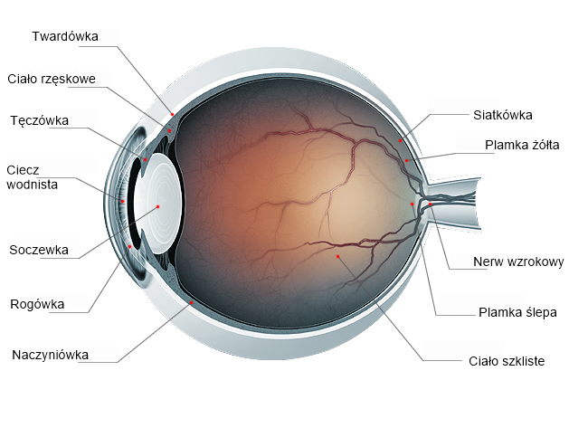
The visual organ consists of the eyeball, which receives visual impressions, the visual pathways that conduct visual stimuli, and the visual centers in the brain cortex where the image is processed. In a healthy eye without vision defects, the eyeball is roughly spherical with a diameter of about 24 millimeters and is located in the front part of the eye socket. It is mostly filled with the vitreous body, a shapeless substance under pressure, which helps the eyeball maintain its spherical shape. The eyeball itself consists of three membranes: the outer fibrous membrane (sclera and cornea), the middle vascular membrane (iris, ciliary body, choroid), and the inner membrane (retina). The outermost layer is the opaque sclera, which in the front part transitions into the transparent cornea. Together, they form a rigid but flexible framework of the eyeball. The cornea is also the main element refracting light in the eye. Directly behind the cornea is the iris, responsible for eye color. The iris has an opening called the pupil, whose width changes depending on light intensity. Between the cornea and the iris is the anterior chamber, filled with a clear fluid.
RETINA AND MACULA LUTEA
The inner membrane is the retina, which receives the image. The retina has a very complex structure. It consists of several layers of cells, including the layer of pigment epithelial cells, photoreceptor cells (rods and cones), as well as nerve and glial cells. Rods are responsible for twilight vision, while cones are responsible for color recognition and daylight vision. In the central part of the retina is the macula lutea, responsible for the most precise vision because it contains the highest concentration of cones. Rods are scattered throughout the retina except in the macula lutea. Nasally from the macula is the optic nerve disc, which lacks light-sensitive cells (is insensitive to light) and is the connection point of light-sensitive cells with the optic nerve.
FUNCTIONS OF THE VISUAL SYSTEM
The visual system has two main functions: it serves as a moving apparatus and a protective apparatus. As a moving apparatus, it moves the eyeball, which is moved by six extraocular muscles. These muscles keep the eyes in a parallel position and enable eye movements. Larger deviations of the eyeball are usually accompanied by head movements. As a protective apparatus, it protects the eye through elements such as the eye sockets, eyelids, eyelashes, conjunctiva, and the tear organ. The eye socket is a hard shield protecting against mechanical injuries. Eyelids and eyelashes protect the eye from excess light, injuries, and impurities and play an important role in moistening the eye. Both eyelids (upper and lower) have eyelashes that help protect the eye especially from various pollens and dust. Eyelids move reflexively and voluntarily. Reflex reactions occur in situations threatening the eye, e.g., when strong wind carries sand or dust, or when the light is too bright. Then we squint by partially closing the eyelids or blink. The latter also plays a role in moistening the eye, as blinking spreads tears, simultaneously cleaning the eye of impurities. The conjunctiva is a transparent mucous membrane lining the inner side of the eyelid and the front surface of the eyeball. The tear organ produces the tear film necessary for the proper functioning of the eyeball. The tear organ consists of the lacrimal gland and drainage pathways: lacrimal canals, lacrimal sac, and nasolacrimal duct. The lacrimal gland lies above the outer corner of the eyelid fissure. It is divided into two parts: a larger orbital part and a smaller palpebral part. The tear drainage pathway begins near the inner corner of the eyelid fissure, where on the posterior edge of each eyelid is the lacrimal papilla, topped by the lacrimal punctum. From each punctum, a lacrimal canal leads to the lacrimal sac. Downward, the lacrimal sac continues as the nasolacrimal duct. The tear film participates in many processes, including dissolving and washing away harmful substances from the eye surface, acting bactericidally and bacteriostatically, participating in corneal metabolism, and maintaining a smooth corneal surface.
MOST COMMON VISUAL ORGAN DISORDERS
| Ailments | Treatment/Therapies | ||
| Inflammation of the cornea, sclera, iris | - Most often, medications are applied to the eye or taken orally. Ignored inflammations can lead to ulcers, perforations, and clouding. Despite treatment, they often tend to recur.-Supportive: NL-Epithalon Therapy | ||
| Retinal detachment | -Retinal detachment can be treated surgically. The course of treatment greatly depends on the time of intervention from the first symptoms. Prompt action allows preservation of vision.-Supportive: NL-Epithalon Therapy | ||
| Macular degeneration | -Treatment depends on the form of the disease. The dry form develops relatively slowly and mildly. It constitutes the vast majority of cases. There is also a wet form, which can lead to significant vision loss within a few days. It is treated with injections of agents that stop disease progression into the eye. The agents used in treating macular degeneration show good effectiveness. They are applied under local anesthesia, so they do not cause pain for patients. -Supportive: NL-Epithalon Therapy | ||
| Glaucoma | -Glaucoma is treated pharmacologically with eye drops, laser, or surgically.-Supportive: NL-Epithalon Therapy | ||
| Cataract | -Cataracts, like glaucoma, are treated with appropriately selected eye drops or by surgical treatment. -Supportive: NL-Epithalon Therapy | ||
RETINITIS PIGMENTOSA
Retinitis pigmentosa, also called pigmentary retinopathy and rod-cone dystrophy, is a hereditary disease from the group of retinal dystrophies. During its development, photoreceptor cells, i.e., light-sensitive elements, undergo atrophy. These include rods located on the retina's periphery, which detect ambient light intensity, enable twilight vision, and allow detection of moving objects, and cones mainly located in the back part of the eyeball, responsible for color detection and daylight vision. Other retinal disorders include age-related macular degeneration and lattice degeneration, often found in people with retinal detachment. The cause of retinal degeneration is inherited faulty genes. Retinitis pigmentosa can be inherited in various ways. If passed on in an autosomal dominant manner, it has a milder course and does not significantly impair vision until the 50s or 60s.
NL-EPITHALON IN PIGMENTARY RETINAL DEGENERATION
According to studies, NL-Epithalon prolongs the functional integrity of the retina in hereditary pigmentary retinitis. Additionally, it improves visual functions in patients with symptoms of vision weakening related to age-related macular degeneration and lattice degeneration often found in people with retinal detachment. The action profile of NL-Epithalon on retinal function involves intensifying the bioelectrical and functional activity of the retina by preserving its morphological structure. Supportive therapy with NL-Epithalon in people with degenerative retinal changes yields a positive clinical effect in 90% of cases. Analysis of NL-Epithalon's action suggests that the tetrapeptide participates in transcription mechanisms common to the base and retina.
NL-EPITHALON IN PROTECTING THE MAINTENANCE OF A PROPERLY FUNCTIONING VISUAL SYSTEM
NL-Epithalon is a strong antioxidant protecting the eyes from the harmful effects of free radicals. It can be supplied to the body by taking it in capsule form. It helps protect the collagen reserves in the cornea, contributing to maintaining its proper condition. It combats all infections within the eyeball, prevents the occurrence of glaucoma and cataracts caused by UV radiation or excessive oxidative stress.
BIBLIOGRAPHY
1.V.Kh Khavinson, Peptides and Ageing. 2002; 3:11-144
2.V.Khavinson, M.Razumovsky, S.Trofimova, R.Grigorian, A.Razumovskaya, Pineal-regulating tetrapeptide epitalon improves eye retina condition in retinitis pigmentosa, 2002; 365-8


NL-GHK-Cu in the nervous system
Administration of NL-PEPTIDES peptides